Uranium »
PDB 1anv-2olb »
1bzo »
Uranium in PDB 1bzo: Three-Dimensional Structure of Prokaryotic Cu,Zn Superoxide Dismutase From P.Leiognathi, Solved By X-Ray Crystallography.
Enzymatic activity of Three-Dimensional Structure of Prokaryotic Cu,Zn Superoxide Dismutase From P.Leiognathi, Solved By X-Ray Crystallography.
All present enzymatic activity of Three-Dimensional Structure of Prokaryotic Cu,Zn Superoxide Dismutase From P.Leiognathi, Solved By X-Ray Crystallography.:
1.15.1.1;
1.15.1.1;
Protein crystallography data
The structure of Three-Dimensional Structure of Prokaryotic Cu,Zn Superoxide Dismutase From P.Leiognathi, Solved By X-Ray Crystallography., PDB code: 1bzo
was solved by
D.Bordo,
D.Matak,
K.Djinovic-Carugo,
C.Rosano,
A.Pesce,
M.Bolognesi,
M.E.Stroppolo,
M.Falconi,
A.Battistoni,
A.Desideri,
with X-Ray Crystallography technique. A brief refinement statistics is given in the table below:
| Resolution Low / High (Å) | 24.70 / 2.10 |
| Space group | H 3 2 |
| Cell size a, b, c (Å), α, β, γ (°) | 86.890, 86.890, 99.010, 90.00, 90.00, 120.00 |
| R / Rfree (%) | 19 / 26 |
Other elements in 1bzo:
The structure of Three-Dimensional Structure of Prokaryotic Cu,Zn Superoxide Dismutase From P.Leiognathi, Solved By X-Ray Crystallography. also contains other interesting chemical elements:
| Copper | (Cu) | 1 atom |
| Zinc | (Zn) | 1 atom |
Uranium Binding Sites:
The binding sites of Uranium atom in the Three-Dimensional Structure of Prokaryotic Cu,Zn Superoxide Dismutase From P.Leiognathi, Solved By X-Ray Crystallography.
(pdb code 1bzo). This binding sites where shown within
5.0 Angstroms radius around Uranium atom.
In total 2 binding sites of Uranium where determined in the Three-Dimensional Structure of Prokaryotic Cu,Zn Superoxide Dismutase From P.Leiognathi, Solved By X-Ray Crystallography., PDB code: 1bzo:
Jump to Uranium binding site number: 1; 2;
In total 2 binding sites of Uranium where determined in the Three-Dimensional Structure of Prokaryotic Cu,Zn Superoxide Dismutase From P.Leiognathi, Solved By X-Ray Crystallography., PDB code: 1bzo:
Jump to Uranium binding site number: 1; 2;
Uranium binding site 1 out of 2 in 1bzo
Go back to
Uranium binding site 1 out
of 2 in the Three-Dimensional Structure of Prokaryotic Cu,Zn Superoxide Dismutase From P.Leiognathi, Solved By X-Ray Crystallography.
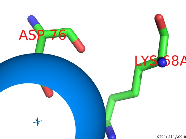
Mono view
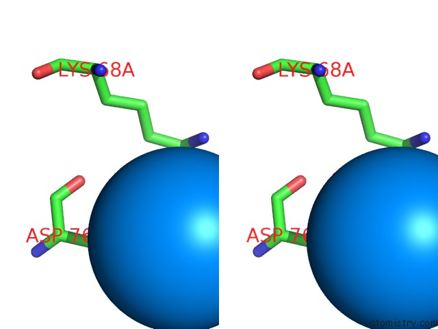
Stereo pair view
Mono view
Stereo pair view
A full contact list of Uranium with other atoms in the U binding
site number 1 of Three-Dimensional Structure of Prokaryotic Cu,Zn Superoxide Dismutase From P.Leiognathi, Solved By X-Ray Crystallography. within 5.0Å range:
|
Uranium binding site 2 out of 2 in 1bzo
Go back to
Uranium binding site 2 out
of 2 in the Three-Dimensional Structure of Prokaryotic Cu,Zn Superoxide Dismutase From P.Leiognathi, Solved By X-Ray Crystallography.
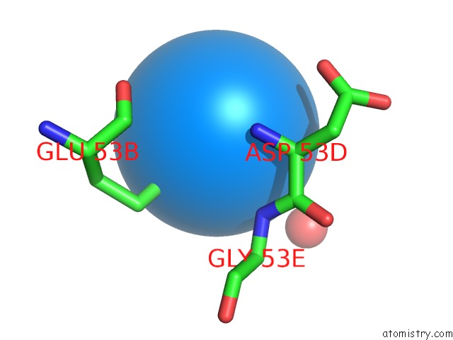
Mono view
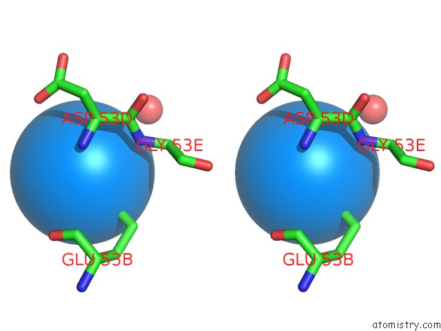
Stereo pair view
Mono view
Stereo pair view
A full contact list of Uranium with other atoms in the U binding
site number 2 of Three-Dimensional Structure of Prokaryotic Cu,Zn Superoxide Dismutase From P.Leiognathi, Solved By X-Ray Crystallography. within 5.0Å range:
|
Reference:
D.Bordo,
D.Matak,
K.Djinovic-Carugo,
C.Rosano,
A.Pesce,
M.Bolognesi,
M.E.Stroppolo,
M.Falconi,
A.Battistoni,
A.Desideri.
Evolutionary Constraints For Dimer Formation in Prokaryotic Cu,Zn Superoxide Dismutase. J.Mol.Biol. V. 285 283 1999.
ISSN: ISSN 0022-2836
PubMed: 9878406
DOI: 10.1006/JMBI.1998.2267
Page generated: Fri Oct 11 10:46:06 2024
ISSN: ISSN 0022-2836
PubMed: 9878406
DOI: 10.1006/JMBI.1998.2267
Last articles
Cl in 7VBVCl in 7V8K
Cl in 7VBU
Cl in 7V73
Cl in 7V68
Cl in 7V57
Cl in 7V46
Cl in 7V44
Cl in 7V43
Cl in 7V3O