Uranium »
PDB 1anv-2olb »
1ncj »
Uranium in PDB 1ncj: N-Cadherin, Two-Domain Fragment
Protein crystallography data
The structure of N-Cadherin, Two-Domain Fragment, PDB code: 1ncj
was solved by
K.Tamura,
W.-S.Shan,
W.A.Hendrickson,
D.R.Colman,
L.Shapiro,
with X-Ray Crystallography technique. A brief refinement statistics is given in the table below:
| Resolution Low / High (Å) | 6.00 / 3.40 |
| Space group | I 4 2 2 |
| Cell size a, b, c (Å), α, β, γ (°) | 99.000, 99.000, 136.100, 90.00, 90.00, 90.00 |
| R / Rfree (%) | 21.2 / 32.1 |
Other elements in 1ncj:
The structure of N-Cadherin, Two-Domain Fragment also contains other interesting chemical elements:
| Calcium | (Ca) | 3 atoms |
Uranium Binding Sites:
The binding sites of Uranium atom in the N-Cadherin, Two-Domain Fragment
(pdb code 1ncj). This binding sites where shown within
5.0 Angstroms radius around Uranium atom.
In total only one binding site of Uranium was determined in the N-Cadherin, Two-Domain Fragment, PDB code: 1ncj:
In total only one binding site of Uranium was determined in the N-Cadherin, Two-Domain Fragment, PDB code: 1ncj:
Uranium binding site 1 out of 1 in 1ncj
Go back to
Uranium binding site 1 out
of 1 in the N-Cadherin, Two-Domain Fragment
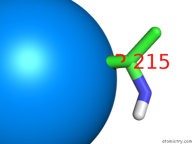
Mono view
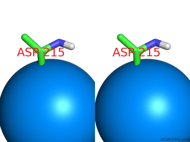
Stereo pair view
Mono view
Stereo pair view
A full contact list of Uranium with other atoms in the U binding
site number 1 of N-Cadherin, Two-Domain Fragment within 5.0Å range:
|
Reference:
K.Tamura,
W.S.Shan,
W.A.Hendrickson,
D.R.Colman,
L.Shapiro.
Structure-Function Analysis of Cell Adhesion By Neural (N-) Cadherin. Neuron V. 20 1153 1998.
ISSN: ISSN 0896-6273
PubMed: 9655503
DOI: 10.1016/S0896-6273(00)80496-1
Page generated: Tue Aug 19 06:54:43 2025
ISSN: ISSN 0896-6273
PubMed: 9655503
DOI: 10.1016/S0896-6273(00)80496-1
Last articles
W in 1DV4W in 1FR3
W in 1GUG
W in 1H9R
W in 1H9K
W in 1H0H
W in 1FEZ
W in 1FKA
W in 1E3P
W in 1E18