Uranium »
PDB 2rkm-7b2w »
3mko »
Uranium in PDB 3mko: Crystal Structure of the Lymphocytic Choriomeningitis Virus Membrane Fusion Glycoprotein GP2 in Its Postfusion Conformation
Protein crystallography data
The structure of Crystal Structure of the Lymphocytic Choriomeningitis Virus Membrane Fusion Glycoprotein GP2 in Its Postfusion Conformation, PDB code: 3mko
was solved by
S.Igonet,
M.C.Vaney,
F.A.Rey,
with X-Ray Crystallography technique. A brief refinement statistics is given in the table below:
| Resolution Low / High (Å) | 37.22 / 1.80 |
| Space group | P 63 2 2 |
| Cell size a, b, c (Å), α, β, γ (°) | 52.925, 52.925, 191.330, 90.00, 90.00, 120.00 |
| R / Rfree (%) | 18.8 / 22.2 |
Other elements in 3mko:
The structure of Crystal Structure of the Lymphocytic Choriomeningitis Virus Membrane Fusion Glycoprotein GP2 in Its Postfusion Conformation also contains other interesting chemical elements:
| Chlorine | (Cl) | 1 atom |
Uranium Binding Sites:
The binding sites of Uranium atom in the Crystal Structure of the Lymphocytic Choriomeningitis Virus Membrane Fusion Glycoprotein GP2 in Its Postfusion Conformation
(pdb code 3mko). This binding sites where shown within
5.0 Angstroms radius around Uranium atom.
In total only one binding site of Uranium was determined in the Crystal Structure of the Lymphocytic Choriomeningitis Virus Membrane Fusion Glycoprotein GP2 in Its Postfusion Conformation, PDB code: 3mko:
In total only one binding site of Uranium was determined in the Crystal Structure of the Lymphocytic Choriomeningitis Virus Membrane Fusion Glycoprotein GP2 in Its Postfusion Conformation, PDB code: 3mko:
Uranium binding site 1 out of 1 in 3mko
Go back to
Uranium binding site 1 out
of 1 in the Crystal Structure of the Lymphocytic Choriomeningitis Virus Membrane Fusion Glycoprotein GP2 in Its Postfusion Conformation
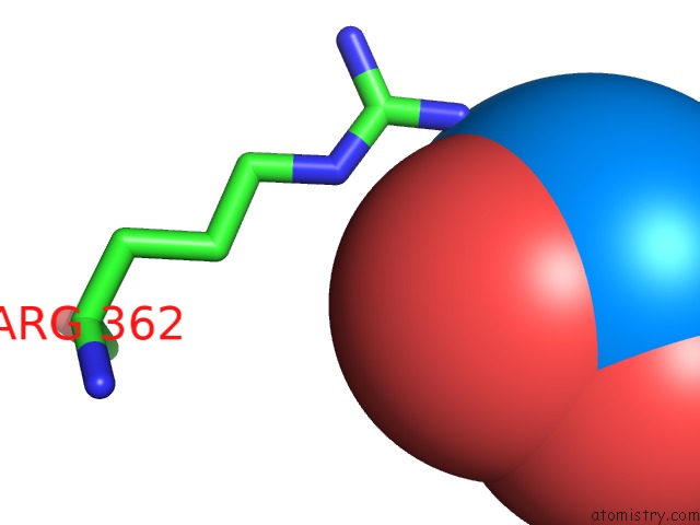
Mono view
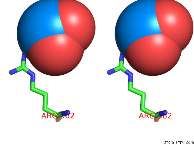
Stereo pair view
Mono view
Stereo pair view
A full contact list of Uranium with other atoms in the U binding
site number 1 of Crystal Structure of the Lymphocytic Choriomeningitis Virus Membrane Fusion Glycoprotein GP2 in Its Postfusion Conformation within 5.0Å range:
|
Reference:
S.Igonet,
M.C.Vaney,
C.Vonhrein,
G.Bricogne,
E.A.Stura,
H.Hengartner,
B.Eschli,
F.A.Rey.
X-Ray Structure of the Arenavirus Glycoprotein GP2 in Its Postfusion Hairpin Conformation Proc.Natl.Acad.Sci.Usa V. 108 19967 2011.
ISSN: ISSN 0027-8424
PubMed: 22123988
DOI: 10.1073/PNAS.1108910108
Page generated: Fri Oct 11 11:01:45 2024
ISSN: ISSN 0027-8424
PubMed: 22123988
DOI: 10.1073/PNAS.1108910108
Last articles
Zn in 9MJ5Zn in 9HNW
Zn in 9G0L
Zn in 9FNE
Zn in 9DZN
Zn in 9E0I
Zn in 9D32
Zn in 9DAK
Zn in 8ZXC
Zn in 8ZUF