Uranium »
PDB 2rkm-7b2w »
4ild »
Uranium in PDB 4ild: Crystal Structure of Truncated Bovine Viral Diarrhea Virus 1 E2 Envelope Protein
Protein crystallography data
The structure of Crystal Structure of Truncated Bovine Viral Diarrhea Virus 1 E2 Envelope Protein, PDB code: 4ild
was solved by
Y.Modis,
Y.Li,
J.Wang,
with X-Ray Crystallography technique. A brief refinement statistics is given in the table below:
| Resolution Low / High (Å) | 47.97 / 3.27 |
| Space group | C 1 2 1 |
| Cell size a, b, c (Å), α, β, γ (°) | 136.720, 54.450, 95.920, 90.00, 92.23, 90.00 |
| R / Rfree (%) | 24.6 / 28.9 |
Other elements in 4ild:
The structure of Crystal Structure of Truncated Bovine Viral Diarrhea Virus 1 E2 Envelope Protein also contains other interesting chemical elements:
| Calcium | (Ca) | 4 atoms |
Uranium Binding Sites:
The binding sites of Uranium atom in the Crystal Structure of Truncated Bovine Viral Diarrhea Virus 1 E2 Envelope Protein
(pdb code 4ild). This binding sites where shown within
5.0 Angstroms radius around Uranium atom.
In total 4 binding sites of Uranium where determined in the Crystal Structure of Truncated Bovine Viral Diarrhea Virus 1 E2 Envelope Protein, PDB code: 4ild:
Jump to Uranium binding site number: 1; 2; 3; 4;
In total 4 binding sites of Uranium where determined in the Crystal Structure of Truncated Bovine Viral Diarrhea Virus 1 E2 Envelope Protein, PDB code: 4ild:
Jump to Uranium binding site number: 1; 2; 3; 4;
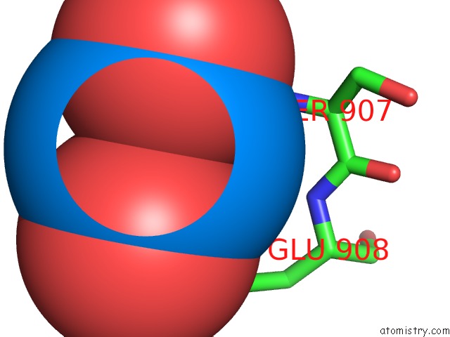
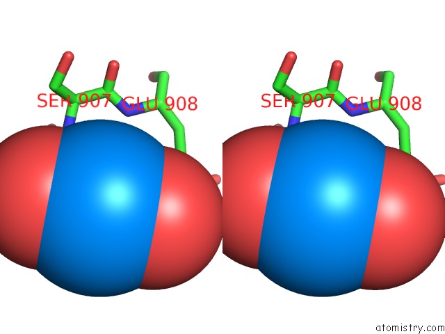
Uranium binding site 1 out of 4 in 4ild
Go back to
Uranium binding site 1 out
of 4 in the Crystal Structure of Truncated Bovine Viral Diarrhea Virus 1 E2 Envelope Protein
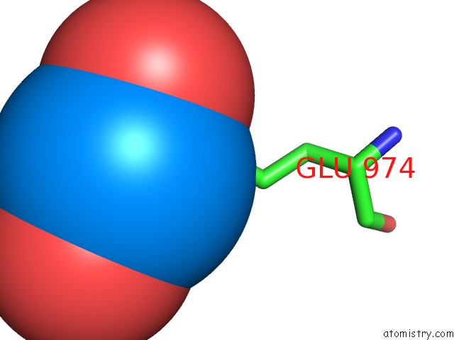
Mono view
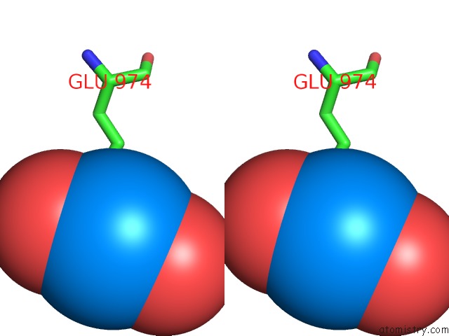
Stereo pair view
Mono view
Stereo pair view
A full contact list of Uranium with other atoms in the U binding
site number 1 of Crystal Structure of Truncated Bovine Viral Diarrhea Virus 1 E2 Envelope Protein within 5.0Å range:
|
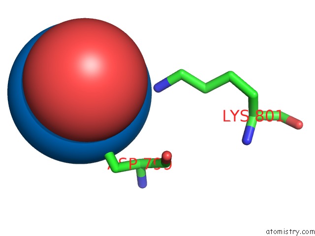
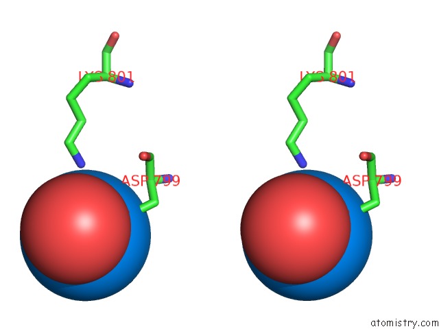
Uranium binding site 2 out of 4 in 4ild
Go back to
Uranium binding site 2 out
of 4 in the Crystal Structure of Truncated Bovine Viral Diarrhea Virus 1 E2 Envelope Protein
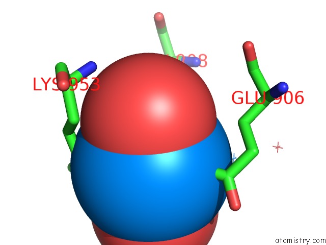
Mono view
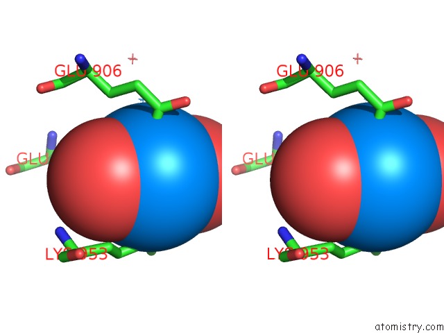
Stereo pair view
Mono view
Stereo pair view
A full contact list of Uranium with other atoms in the U binding
site number 2 of Crystal Structure of Truncated Bovine Viral Diarrhea Virus 1 E2 Envelope Protein within 5.0Å range:
|
Uranium binding site 3 out of 4 in 4ild
Go back to
Uranium binding site 3 out
of 4 in the Crystal Structure of Truncated Bovine Viral Diarrhea Virus 1 E2 Envelope Protein
Mono view
Stereo pair view
Mono view
Stereo pair view
A full contact list of Uranium with other atoms in the U binding
site number 3 of Crystal Structure of Truncated Bovine Viral Diarrhea Virus 1 E2 Envelope Protein within 5.0Å range:
|
Uranium binding site 4 out of 4 in 4ild
Go back to
Uranium binding site 4 out
of 4 in the Crystal Structure of Truncated Bovine Viral Diarrhea Virus 1 E2 Envelope Protein
Mono view
Stereo pair view
Mono view
Stereo pair view
A full contact list of Uranium with other atoms in the U binding
site number 4 of Crystal Structure of Truncated Bovine Viral Diarrhea Virus 1 E2 Envelope Protein within 5.0Å range:
|
Reference:
Y.Li,
J.Wang,
R.Kanai,
Y.Modis.
Crystal Structure of Glycoprotein E2 From Bovine Viral Diarrhea Virus. Proc.Natl.Acad.Sci.Usa V. 110 6805 2013.
ISSN: ISSN 0027-8424
PubMed: 23569276
DOI: 10.1073/PNAS.1300524110
Page generated: Tue Aug 19 07:03:25 2025
ISSN: ISSN 0027-8424
PubMed: 23569276
DOI: 10.1073/PNAS.1300524110
Last articles
W in 1DV4W in 1FR3
W in 1GUG
W in 1H9R
W in 1H9K
W in 1H0H
W in 1FEZ
W in 1FKA
W in 1E3P
W in 1E18