Uranium »
PDB 1anv-2olb »
1ncj »
Uranium in PDB 1ncj: N-Cadherin, Two-Domain Fragment
Protein crystallography data
The structure of N-Cadherin, Two-Domain Fragment, PDB code: 1ncj
was solved by
K.Tamura,
W.-S.Shan,
W.A.Hendrickson,
D.R.Colman,
L.Shapiro,
with X-Ray Crystallography technique. A brief refinement statistics is given in the table below:
| Resolution Low / High (Å) | 6.00 / 3.40 |
| Space group | I 4 2 2 |
| Cell size a, b, c (Å), α, β, γ (°) | 99.000, 99.000, 136.100, 90.00, 90.00, 90.00 |
| R / Rfree (%) | 21.2 / 32.1 |
Other elements in 1ncj:
The structure of N-Cadherin, Two-Domain Fragment also contains other interesting chemical elements:
| Calcium | (Ca) | 3 atoms |
Uranium Binding Sites:
The binding sites of Uranium atom in the N-Cadherin, Two-Domain Fragment
(pdb code 1ncj). This binding sites where shown within
5.0 Angstroms radius around Uranium atom.
In total only one binding site of Uranium was determined in the N-Cadherin, Two-Domain Fragment, PDB code: 1ncj:
In total only one binding site of Uranium was determined in the N-Cadherin, Two-Domain Fragment, PDB code: 1ncj:
Uranium binding site 1 out of 1 in 1ncj
Go back to
Uranium binding site 1 out
of 1 in the N-Cadherin, Two-Domain Fragment
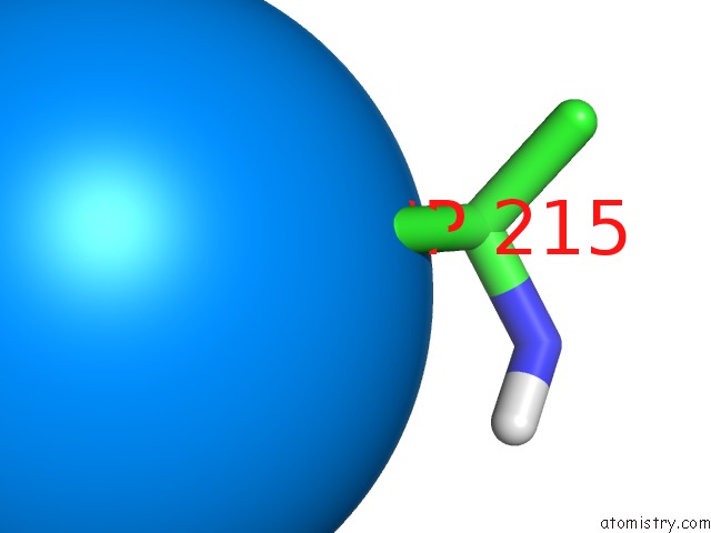
Mono view
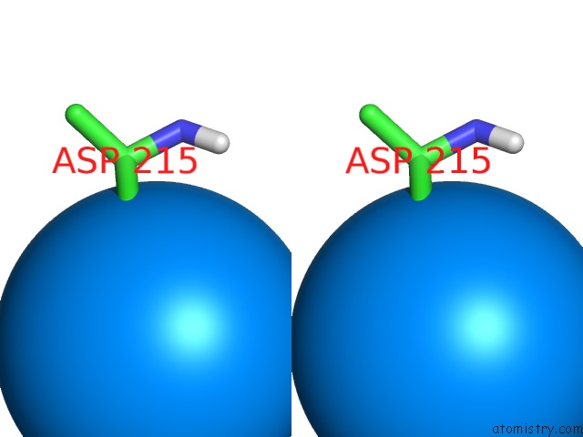
Stereo pair view
Mono view
Stereo pair view
A full contact list of Uranium with other atoms in the U binding
site number 1 of N-Cadherin, Two-Domain Fragment within 5.0Å range:
|
Reference:
K.Tamura,
W.S.Shan,
W.A.Hendrickson,
D.R.Colman,
L.Shapiro.
Structure-Function Analysis of Cell Adhesion By Neural (N-) Cadherin. Neuron V. 20 1153 1998.
ISSN: ISSN 0896-6273
PubMed: 9655503
DOI: 10.1016/S0896-6273(00)80496-1
Page generated: Fri Oct 11 10:51:19 2024
ISSN: ISSN 0896-6273
PubMed: 9655503
DOI: 10.1016/S0896-6273(00)80496-1
Last articles
Zn in 9MJ5Zn in 9HNW
Zn in 9G0L
Zn in 9FNE
Zn in 9DZN
Zn in 9E0I
Zn in 9D32
Zn in 9DAK
Zn in 8ZXC
Zn in 8ZUF